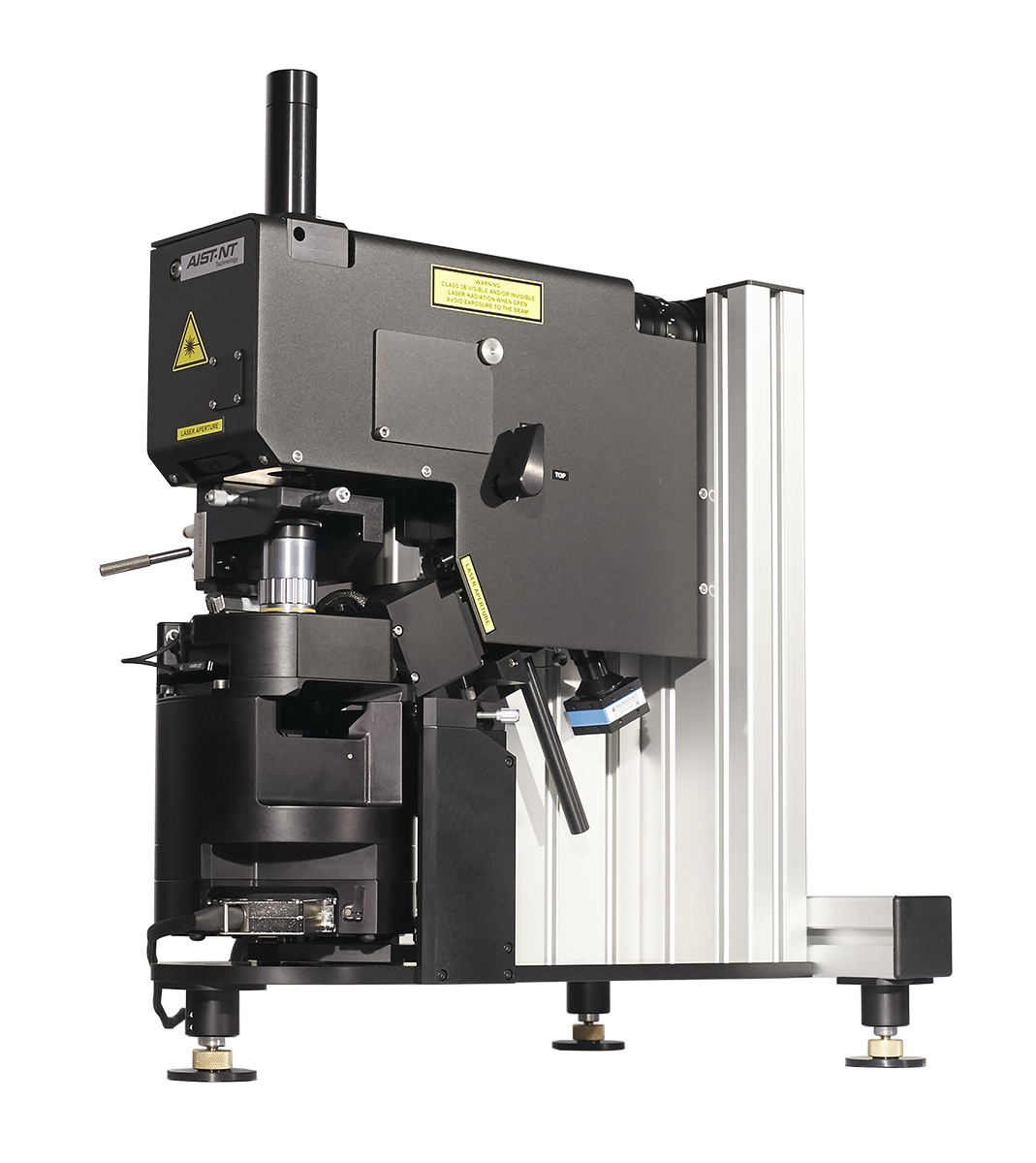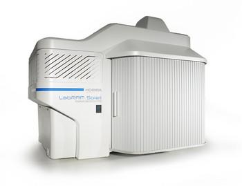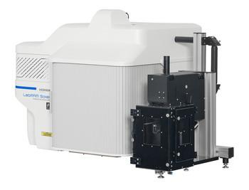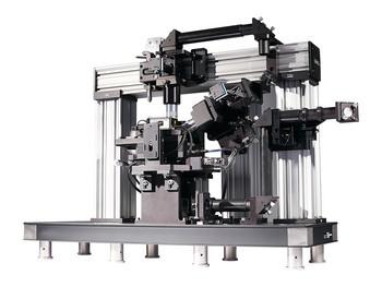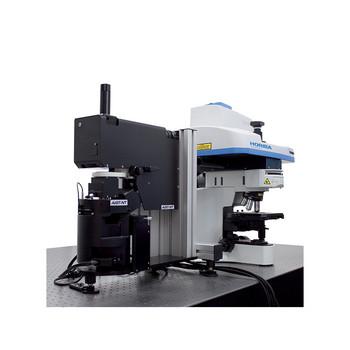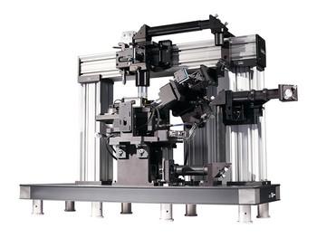
SmartSPM Scanner and Base
Sample scanning range: 100 µm x 100 µm x 15 µm (±10 %)
Scanning type by sample: XY non-linearity 0.05 %; Z non-linearity 0.05 %
Noise: 0.1 nm RMS in XY dimension in 200 Hz bandwidth with capacitance sensors on; 0.02 nm RMS in XY dimension in 100 Hz bandwidth with capacitance sensors off; < 0.04 nm RMS Z capacitance sensor in 1000 Hz bandwidth
Resonance frequency: XY: 7 kHz (unloaded); Z: 15 kHz (unloaded)
X, Y, Z movement: Digital closed loop control for X, Y, Z axes; Motorized Z approach range 18 mm
Sample size: Maximum 40 x 50 mm, 15 mm thickness
Sample positioning: Motorized sample positioning range 5 x 5 mm
Positioning resolution: 1 µm
AFM Head HE002
Laser wavelength: 1300nm;
No registration laser influence on biological sample;
No registration laser influence on photovoltaic measurements;
Registration system noise: <0.1nm;
Fully motorized: 4 stepper motors for cantilever and photodiode automated alignment;
Free access to the probe for additional external manipulators and probes;
Top and side simultaneous optical access: with planapochromat objectives, Side objective up to 100x, NA=0.7 Top objective 10x, NA=0.28 simultaneously;
SPM Measuring Modes
Contact AFM in air/(liquid optional); Semicontact AFM in air/(liquid optional); Non -contact AFM; Phase imaging; Lateral Force Microscopy (LFM); Force Modulation; Conductive AFM (optional); Magnetic Force Microscopy (MFM); Kelvin Probe (Surface Potential Microscopy, SKM, KPFM); Capacitance and Electric Force Microscopy (EFM); Force curve measurement; Piezo Response Force Microscopy (PFM); Nanolithography; Nanomanipulation; STM (optional); Photocurrent Mapping (optional); Volt-ampere characteristic measurements (optional).
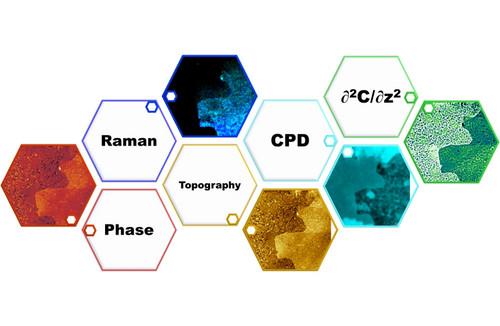
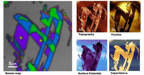
Measuring SPM Modes simultaneously with Raman measurements
Contact AFM in air;
Contact AFM in liquid (optional);
Semicontact AFM in air;
Semicontact AFM in liquid (optional);
Dynamic Force Microscopy (DFM, FM-AFM);
Dissipation Force Microscopy;
True Non-contact AFM;
Phase Imaging;
Lateral Force Microscopy (LFM);
Force Modulation;
Conductive AFM (optional);
Single-pass Kelvin Probe;
Piezo Response Force Microscopy (PFM);
STM (optional);
Photocurrent Mapping (optional);
Shear-force Microscopy with tuning fork (ShFM) (optional);
Normal Force Microscopy with tuning fork (optional).
Spectroscopy Modes
Confocal Raman, Fluorescence and Photoluminescence imaging and spectroscopy
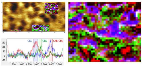
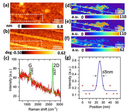
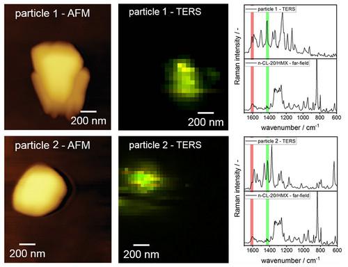
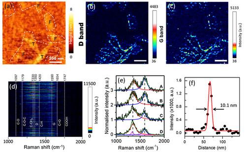
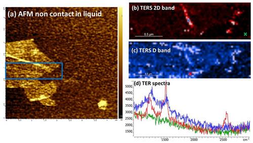
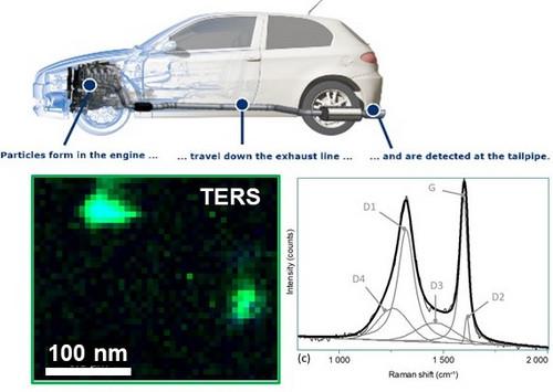
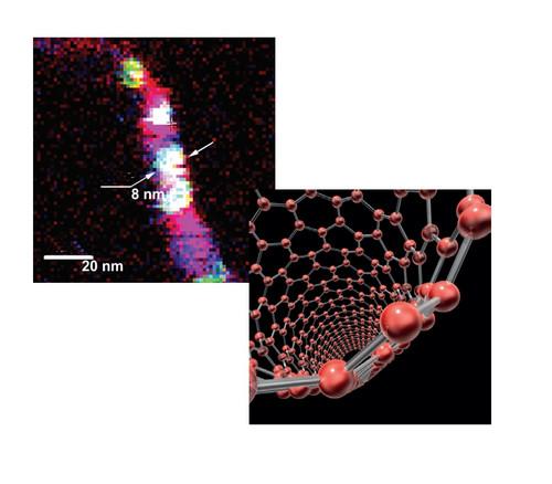
Tip-Enhanced Raman Spectroscopy (TERS) in AFM, STM, and shear force modes
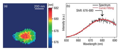
Tip-EnhancedPhotoluminescence (TEPL)
Near-field Optical Scanning Microscopy and Spectroscopy (NSOM/SNOM).
Conductive AFM Unit (optional)
Current range: 100 fA ÷ 10 µA; 3 current ranges (1 nA, 100 nA and 10 µA) switchable from the software.
Optical Access
Capability to use simultaneously top and side plan apochromat objective: Up to 100x, NA = 0.7 from top or side; Up to 20x and 100x simultaneously.
Closed loop piezo objective scanner for ultra stable long term spectroscopic laser alignment: Range 20 µm x 20 µm x 15 µm; Resolution: 1 nm
