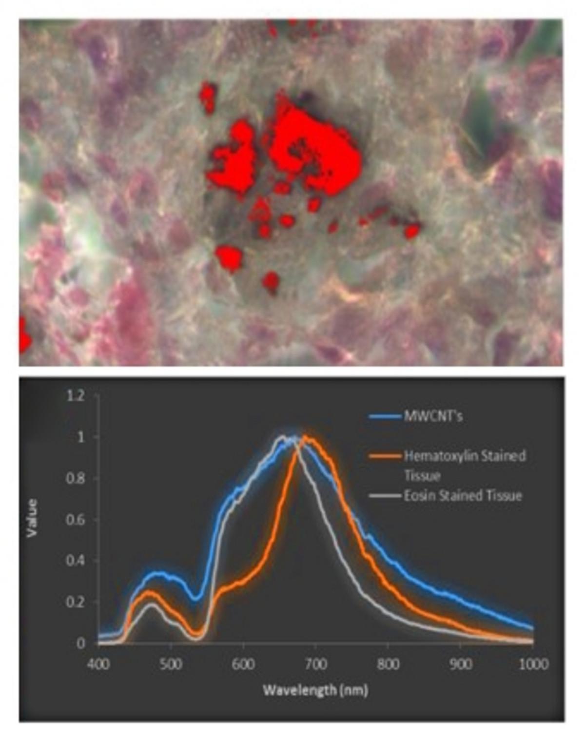

An imaging platform that combines Raman microscopy with enhanced darkfield and hyperspectral imaging technology was used to study lung tissue sections containing carbon nanotubes. It enabled easy identification of the regions containing the carbon nanotubes followed by spectroscopic characterization of chemical composition.
Nanotoxicology requires physico-chemical characterization of nanomaterials in order to examine the health effects of xenobiotic exposure to tissue cultures or model organisms. Such substances can be analyzed with Raman microscopy, which is useful for performing physiological, pharmacological, and toxicological assessments. CytoViva’s patented enhanced darkfield (EDF) illumination is well-suited to detect very small exogenous substances present in complex environments such as tissues with relative ease compared to standard darkfield imaging, followed by hyperspectral imaging (HSI, CytoViva) to differentiate components based on their optical characteristics. Adding EDF and HSI to HORIBA Scientific’s confocal Raman microscope allows the complete analysis of on a single platform (no sample transfer) from detection to differentiation to chemical identification.
MicroRaman Spectrometer - Confocal Raman Microscope
Do you have any questions or requests? Use this form to contact our specialists.
